|
Tests > Diagnostic Tests
Last Update: 03/24/2017
|
Diagnostics | Imaging | Labs | Other Tests
Related Topics
Diagnosis & Pathology | Getting a Second Pathology Evaluation | Prognostic indicators | Performance Standards Diagnostic Tests | Disease Direction Indicators
How Lymphomas are Diagnosed (a concise overview)
Adapted from www.cancer.org
Examination of all biopsy samples by pathologist
The pathologist looks at the appearance, size, and shape of the cells and how the cells are arranged. (sometimes called morphology)
Immunohistochemistry
Biopsy sample is treated with antibodies that attach only to specific molecules on the cell surface. These antibodies cause color changes, which can be seen under a microscope, helping to identify different types of lymphoma and if the cells represent other diseases.
Flow Cytometry to help determine the exact type of lymphoma, or exclude lymphoma
This test also looks for certain molecules on the outside surface of cells by which antibodies (protein molecules) stick, helping to identify what types of cells they are. This test can look at many more cells than immunohistochemistry.
Flow cytometry is most commonly used test for immunophenotyping -- classifying lymphoma cells according to the substances (antigens) on their surfaces. Different types of lymphocytes have different antigens (distinct protein molecules) on their surface. These antigens may also change as each cell matures - goes from one stage of development to another.
Flow cytometry can help determine whether lymph node swelling is due to lymphoma, some other cancer, or a non-cancerous disease (such as reactive hyperplasia - normal immune cells reacting appropriately to an infection).
Cytogenetic tests help identify the type of lymphoma at the gene-level
Evaluation of the chromosomes in the lymphoma cells for abnormalities, such as translocations (wrong order), too many chromosomes (additions), too few chromosomes (deletions).
Molecular Genetic Studies
Tests of the DNA of lymphoma cells to detect abnormalities in cells that can't be seen with a microscope.
Fluorescent in situ hybridization (FISH) iook for specific changes in chromosomes.
FISH can find most translocations that are too small to be seen with usual cytogenetic testing. It uses special fluorescent dyes that only attach to specific parts of chromosomes.
FISH can be used to look for specific changes in chromosomes. It can be used on regular blood or bone marrow samples.
Polymerase Chain Reaction (PCR) can also find translocations too small to be seen under a microscope, even if there are very few lymphoma cells present in a sample.
Can also detect certain genes that have been "turned on" and are contributing to the lymphoma cells' abnormal growth.
Other Lab Tests
While blood tests are not used to diagnose lymphoma, they can be helpful in monitoring for changes in an advanced lymphoma, or to look for abnormalities that warrant further testing.
For example, if the blood counts are low, it might indicate that the lymphoma is growing in the bone marrow and restricting blood cell production. Increasing or decreasing Levels of lactate dehydrogenase or LDH, can provide indications of disease direction or changes in growth tempo.
Other blood tests can help detect liver or kidney problems caused by the spread of lymphoma cells or due to the side effects of treatments.
Blood tests can also help determine if treatment is needed to correct blood levels of certain minerals, or to make sure blood is clotting properly.
.gif) |
technical: Best Practice in Lymphoma Diagnosis and Reporting
British Committee for Standards in Haematology PDF
Royal College of Pathologists
|
|
Biopsy
|
Biopsy
NOTES:
Best to avoid blood thinners prior to having a surgical procedures, such as aspirin, fish oil, Vitamin E. Talk to your doctor about medications and supplements you are taking.
IMPORTANT:
"If sufficient material is available, it is advantageous to snap freeze a small portion of the lymph node in liquid nitrogen. Frozen section immunohistochemistry and DNA gene rearrangement studies can be performed on this material." Bccancer
Return to top
|
A biopsy of suspected lymphoma tissue (typically a lymph node), and subsequent evaluation by a trained pathologist is the only way to definitively diagnose lymphoma. It may take a week or more to get the results, and this time of waiting often causes considerable anxiety for patient and family. The delay is not an indication that a lymphoma was found.
When surgical resection (removal) of a lymph node is not possible, a Fine Needle Aspiration or Large Needle/Core Biopsy may be performed, but each of these procedures have diagnostic limitations. Of the the two, an image-guided core biopsy is more reliable.
|
|
About Biopsy Cancer Help Org | Medline Plus | InteliHealth
|
|
The Biopsy Report: A Patient's Guide - By Edward O. Uthman, MD. Diplomate, American Board of Pathology Biopsy Terms
|
Core Biopsy - Image Guided
|
Effectiveness and Safety of Image-Directed Biopsies Medscape (free login req.) from Southern Medical Journal
|
|
Clinical utility of computed tomography-guided core needle biopsy in the diagnostic re-evaluation of patients with lymphoproliferative disorders and suspected disease progression. Ann Oncol. 2003 Sep;14(9):1438-41. PMID: 12954585
|
Fine Needle Aspiration (FNA)
|
From ajcp.ascpjournals.org:
Fine-Needle Aspiration Cytology in the Diagnosis of Lymphoma The Next Step, by Linda M. Sandhaus, MD http://bit.ly/1aNXroh
|
|
Utilization of fine-needle aspiration cytology and flow cytometry in the diagnosis and subclassification of primary and recurrent lymphoma http://bit.ly/gBXctz
Nancy A. Young M.D.1,*, Tahseen I. Al-Saleem M.D.1, Hormoz Ehya M.D.1, Mitchell R. Smith M.D., Ph.D.2Article first published online: 10 NOV 2000
In conclusion, FNA cytology combined with FC (flow cytometry) obviates the need for tissue biopsy in many instances, particularly when the pathologists have experience diagnosing hematologic malignancies and correlate the findings with the FC results and the clinical history.
Biopsy may be necessary when monoclonality cannot be proven in a clinically suspicious lymph node or when lymphoma grade or subtype cannot be determined.
|
|
Value and limitations of fine-needle aspiration cytology in diagnosis and classification of lymphomas: A review. Diagn Cytopathol. 1999 Oct;21(4):240-9. Review. PMID: 10495316 | Related articles
Lymphomas have successfully been classified by FNA cytology following the prevalent histologic classifications. The success rate of FNA cytology ranges from 80%-90% in diagnosis of NHL and from 67.5%-86% in its subtyping.
The cytodiagnosis of Hodgkin's disease (HD) depends upon demonstration of Reed-Sternberg cells or Hodgkin's cells amongst appropriate reactive cell components. The diagnostic accuracy of FNA cytology for HD has also been invariably high (>85%). Yet, the role of cytology in primary diagnosis, subclassification and management of patients with lymphoma remains controversial. The differential diagnostic problems for NHL include a group of small round cell tumors, nonlymphoid acute leukemias and HD.
Reservations have been expressed regarding the efficacy of cytology in separating florid reactive hyperplasia from low-grade malignant lymphoma.
The reported cytodiagnostic accuracy for follicular lymphomas and nodular sclerosis type of HD is less compared to other subtypes of NHL and HD respectively since nodular pattern and sclerosis are strict histologic criteria which can not be appreciated in cytologic preparations. Entities like atypical lymphoproliferative disorders, peripheral T-cell lymphomas and Ki-1 positive anaplastic large cell lymphomas pose diagnostic challenges to cytologists.
Despite these limitations, FNA cytology remains the first line of investigations (screening test) used in cases of lymphadenopathy. Besides initial diagnosis of lymphoma, it helps in detection of residual disease, recurrences and progression of low-grade to high-grade lymphoma, and helps in staging the disease.
|
|
About Fine Needle Aspiration and Large Needle/Core Biopsy OncologyChannel
"because of small sample sizes and lack of information about lymph node structure, FNA often is inadequate for the initial diagnosis of HD or NHL. In such cases, larger tissue samples are obtained by surgical biopsy." OncologyChannel
|
|
Fine-Needle Aspiration in Non-Hodgkin Lymphoma: Evaluation of Cell Size by Cytomorphology and Flow Cytometry from American Journal of Clinical Pathology - Posted 08/06/2002 Medscape free login req.
|
Image-guided core-needle biopsy (IGNB)
Image-guided core-needle biopsy in patients with suspected or recurrent lymphomas
Abstract
BACKGROUND
It is now commonly admitted that the diagnosis of recurrence of lymphoma can be assessed by image-guided needle biopsy (IGNB). However, the means of obtaining tissue for the original diagnosis of lymphoma is often surgery. The aim of this study was to compare the accuracy of IGNB at the time of diagnosis and at the time of recurrence or progression.
METHODS
The authors performed 212 IGNBs on 194 patients who eventually had a diagnosis of lymphoma. One hundred three IGNBs were obtained at original diagnosis and 109 at recurrence or progression. Large-cutting core-biopsy needles, ranging in size from 20 gauge to 14 gauge, were used. Immunohistochemistry studies were performed in all lymphoma cases.
RESULTS
A diagnosis of lymphoma with subtyping was obtained in 88% of all cases, in 85% at initial diagnosis, and in 89% at follow-up. Therapy was initiated on the basis of IGNB in 93% of all cases, in 91% at initial diagnosis, and in 94% at follow-up. Benign complications occurred in 7.5% of cases and did not require specific treatment. IGNB was equally effective for making a specific diagnosis of lymphoma and initiating therapy at the time of original diagnosis and at follow-up.
CONCLUSIONS
The authors recommend that IGNB be performed as the initial procedure for the diagnosis of lymphoma in the absence of peripheral lymph nodes, either at presentation or at recurrence. Cancer 2000;89:647-52.
Related topics:
|
Recommendations for the Reporting of Lymphoid Neoplasms Medscape (free login req.) A checklist for pathologists and surgeons on how to evaluate and store lymphoid tissue at biopsy.
Why do we need to have this handy? When we ask informed questions, we are more likely to get informed care. Note that it also includes guidance on how to snap freeze the tissue.
|
|
Clinical utility of computed tomography-guided core needle biopsy in the diagnostic re-evaluation of patients with lymphoproliferative disorders and suspected disease progression
http://annonc.oxfordjournals.org/cgi/reprint/14/9/1438.pdf
|
|
Bone Marrow Biopsy
|
|
Moved to new page
|
DiSC assay (optional)
|
|
Return to top
|
Differential Staining Cytotoxicity (DiSC) may be useful to determine what treatment agents will not work on your particular tumor cells.
|
About ACOR.org
|
|
Flow cytometry
|
|
Flow Cytometry Helps to identify the type of lymphoma
An analogy might help.
Like people, lymphocytes have a life span which includes stages of maturation where the appearance changes as do the behaviors and roles in the body as the cells mature.
The type of cell and it's level of maturation is identified by markers - called clusters of differentiation (CD), similar to how people develop features at different ages, (breasts, gray hair, etc)
Specific groups of markers help distinguish between close cousin lymphocytes (follicular, Marginal zone, t-cell, mantle cell, diffuse large b-cell, Hodgkins ...)
The cells are so very small (one billion in a 1 cm lesion) that these differentiation markers cannot be distinguished under a microscope but by "seeing" which fluorescently tagged antibodies stick to the cells.
Return to top
|
Flow cytometry is a way of "measuring the number of cells in a sample, the percentage of live cells in a sample, and certain characteristics of cells, such as size, shape, and the presence of tumor markers (such as Clusters of Differentiation - CD) on the cell surface." NCI
"By virtue of its ability to evaluate not only surface but also cytoplasmic and nuclear antigens, flow cytometry continues to enjoy widespread use in various capacities in lymphoma evaluation and treatment. Additional roles for flow cytometry are likely to be invented in the future and should provide distinctive uses in the molecular era." 4
"It's performed on cells in liquid suspension (i.e. blood, bone marrow, body fluids or tissue cell suspensions) that have been incubated with fluorescently tagged antibodies directed against specific cell surface proteins." ~ Univ. of Washington.
It is "a highly complex process utilized by clinical laboratories to examine blood, body fluids, bone marrow, and tissues. The technology of flow cytometry and the discovery of a method to produce monoclonal antibodies have made possible the clinical use of flow cytometry for the identification of cell populations. This technique measures multiple characteristics within the cell nucleus or cytoplasm (e.g., cell size, internal structure, antigens, DNA, ploidy, and cell cycle analysis) that help distinguish one cell type from another." hgsa.com
-
Flow Cytometry Procedure Manual (Specimen Collections) cancer.gov
-
Flow cytometry in lymphoma diagnosis and prognosis: useful?
Best Pract Res Clin Haematol. 2003 Dec;16(4):583-97. PMID: 14592644
-
Flow cytometric analysis of lymphomas: current status and usefulness.
Arch Pathol Lab Med. 2006 Dec;130(12):1850-8. Review. PMID: 17149963
-
Flow Cytometric Analysis of Leukemia and Lymphoma - The Basics
http://www.med4you
-
Follicular lymphoma differential diagnosis - Stanford
|
FISH
|
|
Return to top
|
 TOPIC SEARCH: Medscape TOPIC SEARCH: Medscape
Interphase fluorescence in situ hybridization (FISH) is an alternative to conventional chromosome analysis of chronic lymphocytic leukemia (CLL) cells.2
The FISH panel is performed for CLL prognosis-specific genomic abnormalities as follows: ATM, Rb-1, D13S25, Trisomy 12, p53 1
-
Chromosome Analysis, Chronic Lymphocytic Leukemia (CLL) Panel by FISH aruplab.com
-
Chronic Lymphocytic Leukemia FISH Panel: Impact on Diagnosis Medscape
|
|
|
TOPIC SEARCH
Medscape
Some terms
Antigens - that which is capable of inducing a specific immune response
Morphology - appearance and structure of cells
Autolysis - spontaneous rupture of cell membrane
Return to top
|
Histology
Histology is the study of cells and tissue on the microscopic level.
Procedures and tests that follow are conducted on tissue obtained from a biopsy.
Frozen Sections
Provides for a rapid provisional diagnosis and the identification of antigens and or enzymes which maybe lost during subsequent fixation and processing schedules. See source for details.
Specimen FixationPreserve the tissue and cells without without distorting or dissolving cellular constituents.
Paraffin SectionsParaffin embedded tissue are cut to a thickness of 3µm. Sections are floated on a warm water bath, then picked up onto microscope slides and allowed to drain. Sections for staining are prepared. See source for details:
Resin SectionsEmbedding tissues in resin offers several advantages over frozen or paraffin sections, such as improved morphology and less shrinkage.
|
|
|
TOPIC SEARCH
Medscape
Some terms
Antigens - that which is capable of inducing a specific immune response
Return to top
|
Immunocytochemistry
The demonstration of antigens in tissue sections or smears by the use of specific immunological (antibody-antigen) interactions culminating in the attachment of a visible marker to the antigen. See source for details.
Source: hmds.org
Immunophenotyping
Reveals the kinds of surface molecules that are present on cells (typically immune cells), such as CD20, CD22.
CD stand for clusters of differentiation, which show the developmental stage of the cell and the cell type. CD20, for example, is expressed only on mature b-cells, but not t-cells.
|
Lumbar puncture / Spinal Tap
(not common)
|
TOPIC SEARCH
Medscape
Return to top
|
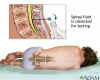 Checks for malignant cells in the cerebrospinal fluid (CSF), which circulates around the brain and spinal cord. Checks for malignant cells in the cerebrospinal fluid (CSF), which circulates around the brain and spinal cord.
|
About CancerHelp UK | Medline Plus | emedicine
|
|
Mediastinoscopy (not common)
|
TOPIC SEARCH
Medscape
Return to top
|
"The mediastinumis is the space that separates the 2 lungs and contains the heart, thymus, esophagus, trachea, the large blood vessels, and lymph nodes. A mediastinoscopy is a procedure in which a lighted instrument (mediastinoscope) is inserted through a neck incision to visually examine the structures in the top of the chest cavity." Source - MedlinePlus
|
About MedlinePlus
|
|
Molecular Diagnostics - LymphoChip
|
TOPIC SEARCH
Medscape *
"Ultimately, this effort will guide patients towards therapies that are tailored for their particular diseases and will identify new molecular targets for therapeutic development."
Return to top
|
"We began a study of gene expression in lymphoid malignancies by constructing a specialized DNA microarray, termed the "Lymphochip", that is enriched in genes which are selectively expressed in lymphocytes and genes which regulate lymphocyte function (1)."
Source: Full text: nih.gov
The LymphoChip promises to:
|
help identify new molecular targets for treatment.
|
|
predict clinical response to therapy and long-term outcome.
|
|
generate gene expression profiles of lymphoma and leukemia.
|
|
help correlate response to treatment with gene expression and thus better match patient to treatment in the future.
|
|
show how treatment affects gene expression.
|
LINKS
|
Application of tissue microarray technology to the study of non-Hodgkin's and Hodgkin's lymphoma. Hum Pathol. 2002 Oct;33(10):968-974. PMID: 12395368 - PubMed
|
|
Molecular Diagnostics
Rita M. Braziel, Margaret A. Shipp, Andrew L. Feldman, Virginia Espina, Mary Winters, Elaine S. Jaffe, Emanuel F. Petricoin III and Lance A. Liotta asheducationbook.org
|
|
PCR testing for
Minimal Residual Disease (MRD)
|
|
Return to top
|
Moved to New Topic page |
Splenectomy
|
|
Return to top
|
"Splenectomy, with an acceptable surgical risk, has the potential to establish the diagnosis of NHL in patients with splenomegaly (enlargement of the spleen) without lymphadenopathy (enlarged lymph nodes) and negative bone marrow findings.
Moreover, splenectomy has the capacity to modify the disease course in patients with NHL complicated by AIHA or hypersplenism."
Source: Splenectomy in patients with malignant non-Hodgkin's lymphoma. Eur J Haematol. 2000 Mar;64(3):145-50. PMID: 10997879 PubMed
|
Serum Free Light Chain Analysis
|
|
Return to top
|
A test for Monoclonal Gamopathy - excess monoclonal immunoglobulin in serum or urine, which indicates excessive plasma cells.
|
Serum Free Light Chain Analysis
http://www.fbr.org/publications/rdlnewsletters/rdlnewsletter_v14n1.pdf |
|
|