What is the
Lymphatic System?
Click to enlarge
Also see Cancer.gov
"Shotty lymph nodes" refers to clusters of small swollen nodes. Shotty nodes may occur when the immune system is reacting to an infection -- it doesn't necessarily point toward any particular disease.
"Bulky disease" generally describes a lymph node or extranodal tumor that measures greater than ten centimeters in any dimension.
calcified nodes are not really rare, and typically are the result of some past, healed infection. TB is well-known to cause them,
for example. Cancer is not."
medhelp.orgs
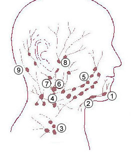
Cervical lymph nodes are lymph nodes located in and around the neck. See Image mage on WebMD Anatomy
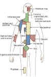
Anatomical Regions for the Staging of Hodgkin Lymphoma
SEER
click to enlarge
Lymphatics - comprehensive list (alphabetical)
http://bit.ly/lymph-alpha
|
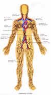
Understanding the lymphatic system
The lymphatic system consists of organs, ducts, and nodes. It transports a watery clear fluid called lymph.
This fluid distributes immune cells and other factors throughout the body. It also interacts with the blood circulatory system to drain fluid from cells and tissues.
The lymphatic system contains immune cells called lymphocytes, which protect the body against antigens (viruses, bacteria, etc.) that invade the body. See more on lymphocytes below. It is abnormal cells of this type that cause lymphoma.
Main functions of the lymphatic system
.gif) |
"to collect and return interstitial fluid, including plasma protein to the blood,
and thus help maintain fluid balance,
|
|
to defend the body against disease by producing lymphocytes,
|
|
to absorb lipids from the intestine and transport them to the blood."
Source jdaross.mcmail.net
|
Also see from this source
Anatomy of lymph system
Role of the lymphatic system in fat absorption and transport
The lymphatic circulation as a drainage system (illustrated)
Lymph organs
Include the bone marrow, lymph nodes, spleen, and thymus. Precursor cells in the bone marrow produce lymphocytes. B-lymphocytes (B-cells) mature in the bone marrow. T-lymphocytes (T-cells) mature in the thymus gland.
Besides providing a home for lymphocytes (B-cells and T-cells), the ducts of the lymphatic system provide transportation for proteins, fats, and other substances in a medium called lymph.
Lymph nodes: "Human lymph nodes are bean-shaped and range in size from a few millimeters to about 1-2 cm in their normal state.
They may become enlarged due to a tumor or infection. White blood cells are located within honeycomb structures of the lymph nodes. Lymph nodes are enlarged when the body is infected due to enhanced production of some cells and division of activated T and B cells.
In some cases they may feel enlarged due to past infections; although one may be healthy, one may still feel them residually enlarged." Wikipedia.org
Lymph
"Means clear water and it is basically the fluid and protein that has been squeezed out of the blood (i.e. blood plasma). The lymph is drained from the tissue in microscopic blind-ended vessels called lymph capillaries.
These lymph capillaries are very permeable, and because they are not pressurized the lymph fluid can drain easily from the tissue into the lymph capillaries.
As with the blood network the lymph vessels form a network throughout the body, unlike the blood the lymph system is a one-way street draining lymph from the tissue and returning it to the blood." Source bbc.co.uk
"Unlike the cardiovascular system, the lymphatic system is not closed and has no central pump." wikipedia.org "Lymph movement occurs despite low pressure due to peristalsis - smooth muscle and skeletal activity (everyday activity and motion of the body).
"Secondary lymphatic tissues control the quality of immune responses. Differences among the various lymphatic tissues significantly affect the form of immunity and relate to how antigens (bacteria, virus, fungus, etc.) are acquired by these organs.
- Lymph nodes are filters of lymph
- the spleen is a filter of blood
- mucosal associated lymphatic tissues acquire antigens by transcytosis to lymphoid tissue from the "external" environment across specialized follicle-associated epithelial cells." Source geocities.com
"Lymphatics are found in every part of the body except the central nervous system. The major parts of the system are the bone marrow, spleen, thymus gland, lymph nodes, and the tonsils. Other organs, including the heart, lungs, intestines, liver, and skin also contain lymphatic tissue." gorhams.dk
Lymphoma
Is a disease in which malignant lymphocytes grow too fast or live too long. These cells may then accumulate in the lymph nodes or other areas of the lymphatic system to form tumors. When these cells accumulate in lymph nodes it's often called adenopathy - the enlargement of the lymph nodes; but adenopathy can have other causes.
There are many benign reasons for enlarged lymph nodes:
Diagnostic Problems in the Evaluation of Lymphadenopathy, Weinstock, Straus Cancernetwork.com
Lymphadenopathy: Differential Diagnosis and Evaluation, Robert Ferrer, M.D., M.P.H. American Family Physician
Normal sized lymph nodes?
Human lymph nodes are bean-shaped. These organs of the immune system can range in size from a few millimeters to about 1–2 cm in their normal state. They may become enlarged due to a tumor or infection, or lymphoma.
Warwick, Roger; Peter L. Williams (1973) [1858]. "Angiology (Chapter 6)". Gray's anatomy.
|
Swollen lymph nodes By Mayo Clinic staff
|
Reactive Lymph nodes
"LYMPH nodes are a combination of burglar alarm and West Point. Like a burglar alarm they are on guard against intrusive antigen. Like West Point, the nodes are in the business of training a militant elite:" pleiad.umdnj.edu
What's the deal with Waxing, Waning, and Persistently Enlarged Lymph Nodes?
Lymph nodes can increase or decrease in size for many reasons, including response to treatment, progression of lymphoma, spontaneous regression of lymphoma, immune activation against lymphoma, infection or the resolving of infection (reactive, see above), and so on.
Therefore, imaging of lymphoma is only an estimate of treatment response and disease direction.
|
Collapsing of necrotic areas in lymph nodes may explain a sudden decrease in a large lymph node.
|
|
Residual lymphoid masses?
Following treatment "a residual mass persisting on CT after treatment poses a common clinical dilemma: it may indicate the presence of viable lymphoma, which requires further treatment, or it can be benign, consisting of only fibrotic and necrotic tissues." PMID: 12644887
For this reason PET or Gallium scans may be used after treatment to help differentiate active disease from scar tissue.
Lymphoid tumors are made up of the accumulated abnormal lymphocytes but also supportive tissue of different cell types: “epithelial cells and also there is supporting tissue, called connective tissue which is there to support the epithelial cells”. These cells are sometimes referred to in shorthand as tumor “stromal” cells.
Our understanding is that the macrophages (immune cells) will eventually gobble up this necrotic material after successful therapy, which explains why the treated “tumor” continues to shrink well after therapy is done.
NOTE: The delayed shrinking of lymphoid tumor can lead to questionable conclusions of the causal effect of subsequent therapy – standard, investigational, and alternative, because the tumor in some cases would have resolved with more time.
PET scans are used to help distinguish between necrotic and viable tumor following treatment, but this test is not perfect … can produce false positives due to inflammation.
|
|
Reactive lymph nodes - Lymph nodes may enlarge when immune cells react to pathogen such as virus or bacteria. Swollen glands, common to many illnesses is an example of nodes enlarging in response to a pathogen. This might also be called a flare.
|
Resources
|
Blood Cells www.siumed.edu
|
|
Lymphatic System bartleby.com | gorhams.dk | cancerhelp.org.uk
|
|
Lymphedema PAL
|
|
Lymphatics - comprehensive list (alphabetical) http://bit.ly/lymph-alpha
|
|
Quiz: Quiz: Know your nodes: knowyournodes.org
Leukemia and Blood foundation: Now is the time to get to ‘know your nodes’, and find out just how important your lymphatic system is.
|
|
Table of Lymphatic vessels (comprehensive) anatomy.uams.edu
|
|
Spontaneous regressions PAL
|
|
Imaging residual masses: Gallium-67 scintigraphy: a cornerstone in functional imaging of lymphoma.
Eur J Nucl Med Mol Imaging. 2003 Jun;30 Suppl 1:S65-81. Epub 2003 Mar 18. Review PMID: 12644887
|
|
Lymphocytes
Return to top
|
Lymphocytes, also called white blood cells, are described below. These are small cells, 7-9 µm in diameter in blood smears ...
These cells are the second most common white blood cell type, comprising about 30 % of the leukocyte population in peripheral blood....
Lymphocytes travel in the blood, but they routinely leave capillaries and wander through connective tissue. Therefore, lymphocytes may be normally encountered at any time in any location. They even enter epithelial tissue, crawling between the epithelial cells. They reenter circulation via lymphatic system channels (hence their name)."
Source: Blood Cells www.siumed.edu
Life cycle of lymphocytes
|
Develop in the thymus gland or bone marrow.
|
|
B-cells grow (differentiate) and mature in the bone marrow.
|
|
T-cells also start out in the bone marrow. They differentiate and mature in the thymus gland (beneath breastbone).
|
|
B-cell and T-cell lymphocytes are distributed through the blood stream, which eventually branches into tiny blood vessels called capillaries.
|
|
Some lymphocytes migrate to capillaries into surrounding tissues. Some enter lymphatic vessels-tiny, blind-ended tubes-and lead to larger lymphatic ducts and branches.
|
|
Along the way, the fluid passes through lymph nodes, oval structures composed of lymph vessels, connective tissue, and white blood cells. Here, the lymphocytes either are filtered out or are added to the contents of the node.
|
|
|

Click to enlarge
Return to top
|
Lymph nodes: "Human lymph nodes are bean-shaped and range in size from a few millimeters to about 1-2 cm (10 to 20 mm) in their normal state.
They may become enlarged due to a tumor or infection. White blood cells are located within honeycomb structures of the lymph nodes. Lymph nodes are enlarged when the body is infected due to enhanced production of some cells and division of activated T and B cells.
In some cases they may feel enlarged due to past infections; although one may be healthy, one may still feel them residually enlarged." Wikipedia.org
The lymph node provides an environment where lymphocytes can receive initial exposure to foreign antigens (viruses, bacteria, fungi, etc.), which activates the lymphocytes to perform immune functions.
Most lymph nodes form in clusters throughout the system, such as in the neck, armpit, and groin.
|

![[Illustration]](spleen_small.gif)